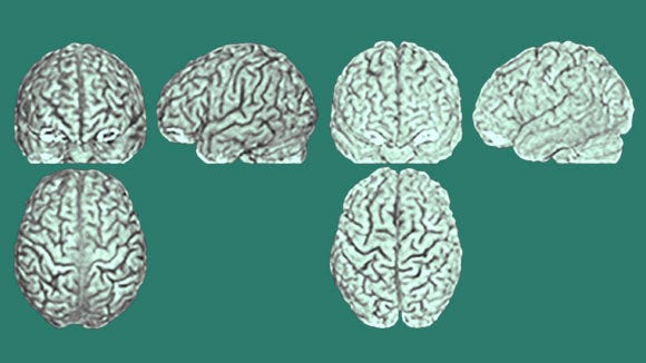
How We Can Compare Locations in People's Brains, Despite Differences in Size and Shape
The answer: brain atlases.
Have you ever wondered how neuroscientists find out what brain regions participate in doing what tasks when people’s brains differ in size, shape, and folding pattern?

Human brains vary in length, width, height, volume, and weight. Age, biological sex, country of origin, specific genes, and experiences, such as playing an instrument—all affect these measures.
People’s brains vary so much that you can actually identify individuals by features of their neuroanatomy.
So, if you flattened out everyone's brains, neither the outline nor the anatomical landmarks within would "line up."
If the equivalent clumps of brain tissue aren’t located in exactly the same place in each person, how do we know how whether they’re more active than the rest of the brain at a given time?
(Remember, the whole brain is always doing something, even when we're just awake and at rest, so there’s a baseline level of activity. So, when we want to know what parts of the brain are doing a task, we’re looking for a big increase).
If the brains being compared don't quite line up, the blobs or "areas of activation" would have fuzzy edges. Because bits of tissue are active in some participants and not others, there’d be less certainty about which are activated during a task and which are not.
Neuroscientists solve this problem by using a brain atlas.
How Brain Atlases Work
A brain atlas is a coordinate system for comparing human brains in 3D space. First, it first divides space into 3 dimensions, x, y, and z. These correspond to the length, height, and depth of the brain.
Then, one or more "normal" human brains are chosen as the “template,” or standard brain.
Through specific mathematical processes, each participant’s brain scan is adjusted to make it match the standard brain(s).
Thus, researchers are really asking where the brain is activating if everyone's brain were the same size and shape.
And fortunately, that works well enough. Amazingly, people's brains are similar enough to be adjusted without wild distortion.
Setting the Standard
You might be wondering, where does the standard brain come from? Whose was it? How was it chosen?
In fact, there’s more than one standard brain, because there are multiple brain atlases: researchers frequently use as many as 9.
The number of people whose brains were scanned to create these brain atlases ranges from 1 to 305.
The most frequently used atlases are the Talairach (pronounced ta-luh-RACK) and the MNI, which stands for Montreal Neurological Institute.
The Talairach and Tournoux atlas, created in 1988, introduced the essential ingredients for modern brain atlases:
A coordinate system. (Theirs was based on 2 anatomical landmarks, including the posterior commissure (PC) and the anterior commissure (AC), which was defined as the origin (0) point.
A template: A brain mapped onto the coordinate system, to be used as a “standard.”
A mathematical transformation to match any brain you study to the template.

Image showing the coordinate axes in relation to the whole brain and the head, from Brett, Johnsrude’s, and Owen’s 2002 discussion of “the problem of functional localization in the human brain,” meaning, the problem of determining which areas in the brain do what.

This pioneering atlas became one of the standards. However, it had a few problems:
It was based on only 1 brain: the postmortem brain of 1 60-year-old French woman.
It used only 2 dimensions.
It did not cover the entire brain space.
So, in 1995, the Montreal Neurological Institute (MNI) created a new atlas to address these problems, the MNI-305, by averaging 3D brain images from 305 living, right-handed people. They were younger and probably healthier than the woman from Talairach and Tournoux’s atlas, with an average age of 23 years.
Most statistical analysis software packages, and the International Consortium for Brain Mapping (ICBM), use a 2001 MNI update, the MNI-152. It adapts the Talairach-Tornoux coordinate system into 3D space. Based on MRI images from 152 “normal” people, it covers the entire brain and cerebellum.
The More Atlases, the Merrier
More recently, various countries have made their own brain atlases to better represent their own populations. Because, as it turns out, the “average” length, width, and height — and the position of various anatomical landmarks — varies across countries.
The Chinese template uses MRI images from 56 right-handed males, with an average age of 24.
The Korean brain template (2005) uses MRI and PET images from 78 “normal,” right-handed Koreans, ages 18-77.
The creators also grouped participants by age (<55, >55) and sex (male, female). These subgroups matter because older brains are thinner and lighter than younger brains, while male brains on average are larger and heavier than female brains. Controlling these variables makes for cleaner results.
A French template comes from a different approach: using multiple MRI scans from 1 person.
International research already suffers from the assumption that all human minds work the same as so-called WEIRD minds (Western, Educated, Industrialized, Rich, and Democratic). We don't want to make that mistake with neuroanatomy by literally reshaping other people’s brains into WEIRD brains.
In general, the brain atlas chosen for a study should match the population being investigated as closely as possible—in country of origin, age, sex, and any health conditions present. That minimizes the amount of distortion of each participant’s brain needed to get useful information.
The Near Future of Brain Atlases
To explain the idea of a “brain atlas,” I’ve discussed ones based on MRI images — the kind I myself have used. These atlases let us examine the relationship between a specific spatial location, its anatomical structure, and its function.
Atlases can also be made from other types of data to answer other questions.
You can use extra high-resolution data to look at smaller spatial-area “micro-circuitry.” The Big Brain and Blue Brain projects focus on micro-circuitry, while several other projects include templates at both micro- and macro-scales.
You can introduce the 4th dimension, time, to create a standard atlas of brain development or aging, as in the Chinese Color Nest Project. There’s even a database of MRI template brains, measured from 2 weeks through 89 years of age. That means age-specific standard brains for almost the entire human lifespan!
You could use genetic data to map where genes are expressed in the brain, as in the Allen Brain Atlas.
You can map connectivity patterns. Connectivity is the relationship between structure or activity in multiple parts of the brain, which form a network or circuit. These occur at both microscale and macroscale. Connectivity analysis lets us look at how a whole network might relate to behavior. The Human Connectome, Brainnetome, and Chinese Color Nest projects all address this question.
You can compare maps for typical vs atypical brains in any of these measures (as in the Japanese Brain/MINDS project).
Ever more types of brain atlases are appearing to address ever more ambitious questions about the relationships between genes, the brain, typical and atypical development, and behavior.
Now, groups are finding ways to integrate data from multiple brain atlases. You can see an example of an interactive, multilevel human brain atlas on the Human Brain Project’s website.
Watch for news about studies integrating different types of brain atlases and data to draw new connections between genes, brain structure at different levels, connectivity, development, and population…or to compare ever more finely-drawn populations.
(You can find out what brain atlas(es) researchers used for a study by skimming the Methods section).
Remember…
Next time you read a neuroimaging study that talks about brain activity in particular locations, remember the results come with a disclaimer: "to the extent every person's brain is the same."
Were you surprised how different even identical twins’ brains look?
Did it surprise you that for much of neuroimaging’s history, one of the top 2 brain atlases was based on the brain of just 1 person?
Any other thoughts, questions, or experiences to share? Was something confusing? Did I get a fact wrong? Comment below; I’d love to hear.


